Digestive / VISUAL EXPLAINER
Where Does Facial Skin Become the Inner Lining of Your Lip?
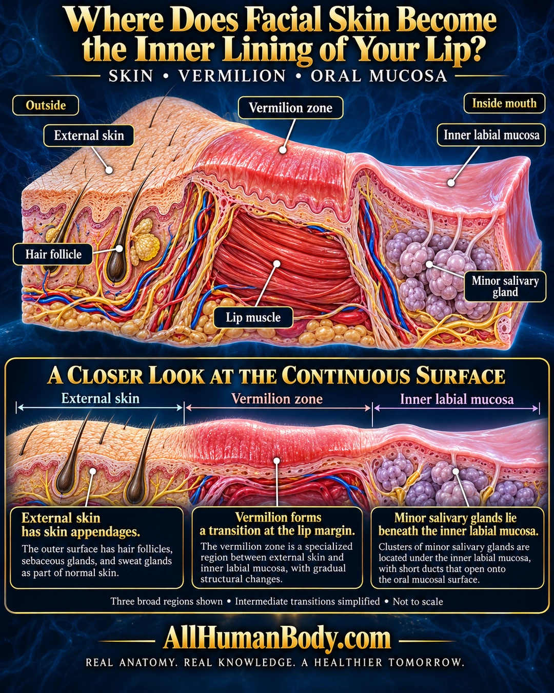
The lip’s surface transitions from external skin through vermilion to inner oral mucosa. Hair-bearing skin and minor salivary glands occupy different regions along that continuous surface.
READ THE IMAGE
What the illustration shows
English poster on navy shows an original unfolded lower-lip surface cutaway from outside left to inside mouth right. Hair-bearing peach external skin transitions across a coral vermilion region into smooth pink inner labial mucosa. Hair follicle pointers land only externally. Purple lobules under inner mucosa are labeled minor salivary gland, with short ducts toward the oral surface. Red lip muscle lies beneath the central region. Lower continuous strip repeats the three broad surface regions with descriptive text. Qualifier notes intermediate transitions simplified and not to scale. Gold AllHumanBody.com and full slogan below.
How to interpret this poster
- Three broad regions simplify intermediate transitions. This is not a literal section shape, universal pigmentation guide or precise epithelial thickness map.
- AI-assisted educational illustration; not physician certification, microscopy, a diagnosis or individualized medical advice.
- Fine annotations need enlargement; the full description provides an accessible text alternative.
- Display the full uncropped illustration; do not trim titles, legends or boundary annotations.
AI-assisted educational illustration, adapted into web-sized images without changing its composition. This is general education, not an individual diagnosis or treatment recommendation.
EVIDENCE / CONTEXT
Sources and what they support
- University of Leeds oral histology ↗
Whole-lip section identifies hairs, vermilion, oral mucosa, muscle and accessory glands.
- Primary human cadaver histology study, 31 lower lips ↗
Distinct epidermal, vermilion, intermediate and labial mucosal differentiation. Use three broad regions with intermediate transition acknowledged, not an absolute three-zone histology claim.
Evidence correspondence is limited to the abstract.