Digestive / VISUAL EXPLAINER
Is the Esophagus Made of Just One Kind of Muscle?
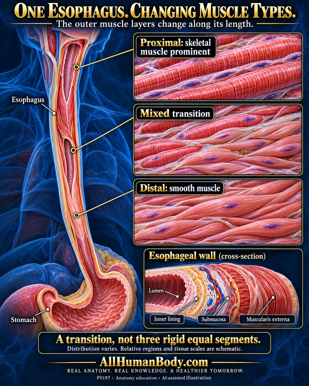
The esophageal muscularis externa changes along its length: skeletal muscle is prominent proximally, a mixed transition follows, and smooth muscle predominates distally. This concerns the propulsive muscle coat, not the epithelial lining or three rigid equal segments.
READ THE IMAGE
What the illustration shows
Gold-titled medical poster on deep navy with a translucent blue neck and chest. On the left an opened esophagus continues down to a stomach. Three gold leaders link relative proximal, transition and distal locations to right-side tissue windows: striated red skeletal fibers with purple peripheral nuclei; a mixture of striated fibers and smooth spindle cells; and unstriated smooth spindle cells. A lower wall cross-section labels the lumen, inner lining, submucosa and outer muscularis externa. The takeaway says the transition is not three rigid equal segments. The gold AllHumanBody.com brand and full slogan sit on a plain navy footer. Opened views, tissue scales and regional proportions are illustrative, not an exact histological or measured distribution map.
How to interpret this poster
- Relative regions, window positions, wall thickness and magnifications are illustrative, not measured proportions or exact histology.
- The supporting human study examined11 cadavers; its distribution is not a universal population norm or fixed boundary.
- Muscle type does not establish a voluntary-control switch. No reflux or swallowing-disorder advice.
- AI-assisted educational illustration; not physician certification, microscopy, a diagnosis or individualized medical advice.
- Fine annotations need enlargement; the full description provides an accessible text alternative.
- Display the full uncropped illustration; do not trim titles, legends or boundary annotations.
AI-assisted educational illustration, adapted into web-sized images without changing its composition. This is general education, not an individual diagnosis or treatment recommendation.
EVIDENCE / CONTEXT
Sources and what they support
- University of Leeds Histology: Oesophagus ↗
Outer muscle transition and distinction from mucosa/submucosa.
- Muscle anatomy of the human esophagus ↗
Eleven-cadaver anatomy study challenges rigid equal-thirds descriptions; no population percentage adopted.
Evidence correspondence is limited to the abstract.