Digestive / VISUAL EXPLAINER
Which Parts of a Tooth Contain Living Tissue?
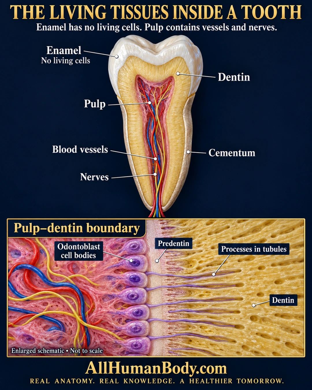
A tooth contains distinct tissues: mature enamel has no living cells, while the central pulp contains vessels and nerves. Odontoblast bodies lie at the pulp border and extend processes into dentinal tubules; cementum covers the root.
READ THE IMAGE
What the illustration shows
Portrait anatomy poster on a navy background titled “The Living Tissues Inside a Tooth.” A large tooth cutaway labels the white enamel cap, yellow dentin, red central pulp, blood vessels, nerves and thin cementum at the root. The subtitle states that enamel has no living cells and pulp contains vessels and nerves. Below, an enlarged schematic of the pulp–dentin boundary labels purple odontoblast cell bodies, a pale predentin band and slender purple processes extending into tubules in the golden dentin. Red and blue vessels and yellow nerves remain on the pulp side. The inset says “Enlarged schematic • Not to scale.” Gold AllHumanBody.com branding and the complete project slogan appear at the bottom.
How to interpret this poster
- Representative tooth form, not a named-tooth reconstruction or patient scan.
- Process lengths, cell sizes and tissue thicknesses are schematic; processes need not fill every tubule completely.
- No pain-transmission, regeneration, repair or dental-treatment claim.
- AI-assisted educational illustration; not physician certification, microscopy, a diagnosis or individualized medical advice.
- Fine annotations need enlargement; the full description provides an accessible text alternative.
- Display the full uncropped illustration; do not trim titles, legends or boundary annotations.
AI-assisted educational illustration, adapted into web-sized images without changing its composition. This is general education, not an individual diagnosis or treatment recommendation.
EVIDENCE / CONTEXT
Sources and what they support
- American Dental Association MouthHealthy: Tooth ↗
Enamel, dentin, cementum and living vascular/innervated pulp.
- University of Leeds Histology Guide: Mouth, lip and teeth ↗
Odontoblast/predentin boundary and processes in dentin.