Nervous / VISUAL EXPLAINER
How Do Signals Enter and Leave Your Spinal Cord?
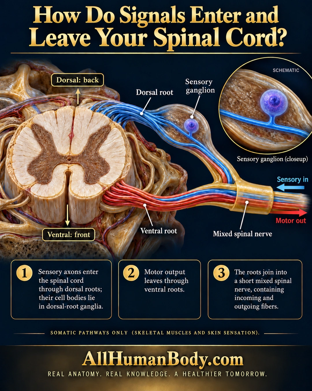
Sensory axons enter spinal-cord context through dorsal roots, with their cell bodies in dorsal-root ganglia. Efferent fibers leave through ventral roots, and the roots join a short mixed spinal nerve; this picture selects somatic pathways.
READ THE IMAGE
What the illustration shows
English poster titled “How Do Signals Enter and Leave Your Spinal Cord?” A dimensional spinal-cord cross-section has dorsal/back above and ventral/front below. Blue sensory fibers run through a labeled dorsal root and sensory ganglion; red motor fibers leave through a separate ventral root. The two roots join into a mixed spinal nerve. A blue arrow points toward the cord and a red arrow away. A circular schematic enlarges a round sensory-cell body connected by one short stalk to an axon. Three text panels explain sensory entry, motor exit and the mixed nerve, with a note limiting the diagram to somatic pathways. Gold AllHumanBody.com branding and the project slogan appear below.
How to interpret this poster
- Autonomic efferents also use ventral roots at applicable levels; this is not an all-efferents-are-somatic claim.
- No rami, exact vertebral level, dermatome map or complete meningeal anatomy is shown.
- The one-cell ganglion inset is schematic, not a relay synapse or a cell-count claim.
- AI-assisted educational illustration; not physician certification, microscopy, a diagnosis or individualized medical advice.
- Fine annotations need enlargement; the full description provides an accessible text alternative.
- Display the full uncropped illustration; do not trim titles, legends or boundary annotations.
AI-assisted educational illustration, adapted into web-sized images without changing its composition. This is general education, not an individual diagnosis or treatment recommendation.
EVIDENCE / CONTEXT
Sources and what they support
- Neuroscience Online: Anatomy of the Spinal Cord ↗
Segmental root direction and joining into a mixed spinal nerve.
- Anatomy and Physiology 2e: 13.4 The Peripheral Nervous System ↗
Sensory ganglia and spinal sensory/motor relationships.