Nervous / VISUAL EXPLAINER
Where Do Purkinje Cells Sit in the Cerebellum?
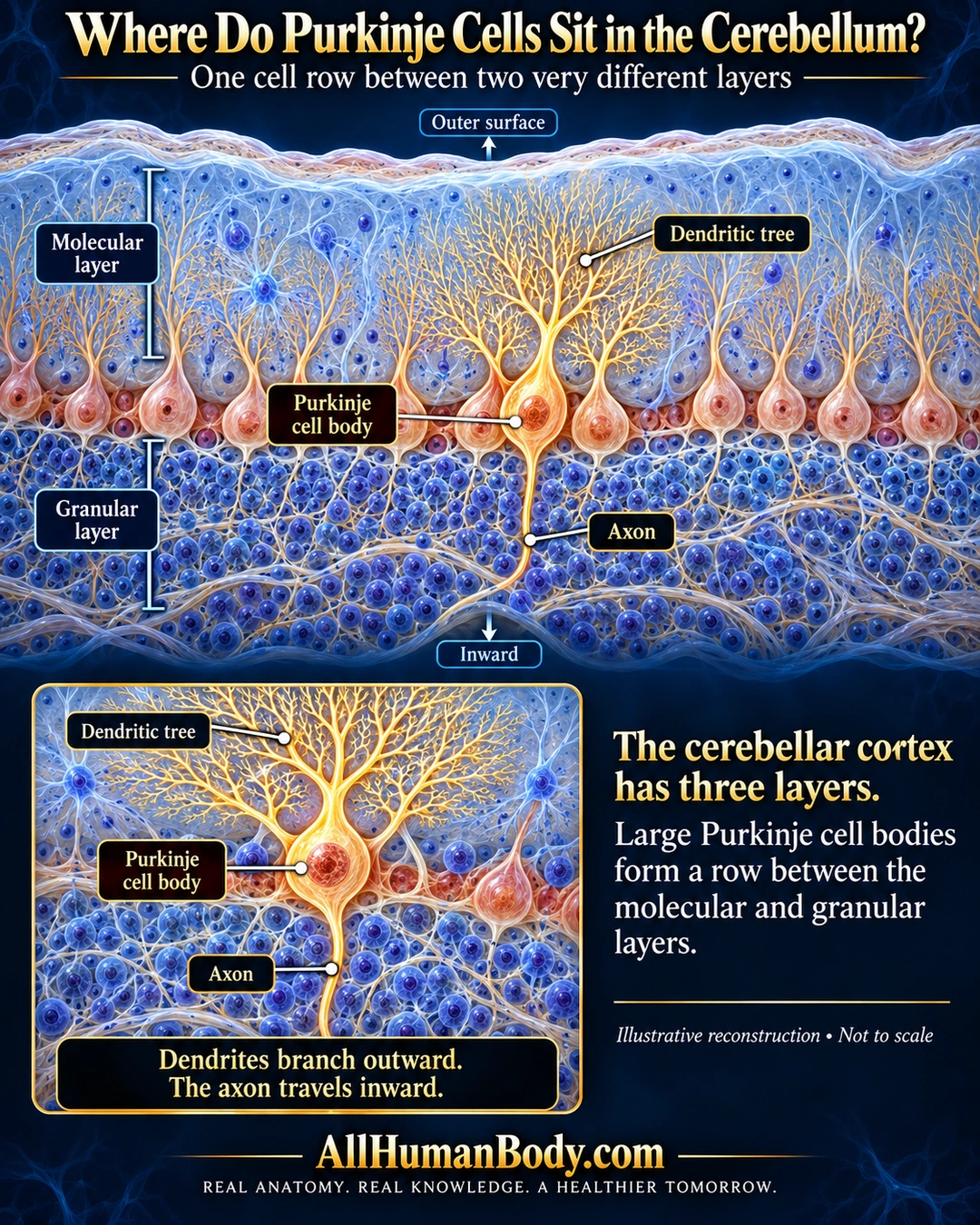
The cerebellar cortex has molecular, Purkinje-cell and granular layers. Large Purkinje cell bodies form a row between the other two layers, with dendrites extending into the molecular layer and an axon passing inward through the granular layer.
READ THE IMAGE
What the illustration shows
English medical poster showing a reconstructed strip of cerebellar cortex on navy blue. A pale-blue molecular layer lies above a single row of peach Purkinje cell bodies, with densely packed blue granular cells below. A selected gold cell sends a branching dendritic tree upward and a slender axon downward through the granular layer. Labels identify layers, cell body, dendritic tree and axon. A lower inset repeats this relationship. Gold AllHumanBody.com branding and the full slogan appear at the foot.
How to interpret this poster
- Illustrative reconstruction, not a single thin stained section, exhaustive circuit or measured cell count.
- The molecular layer is not cell-free; granular-layer cells are not all Purkinje somata. Branch complexity, proportions and glow are schematic.
- Cerebellar cortex is not six-layer cerebral cortex, and these neurons are not cardiac Purkinje fibers.
- AI-assisted educational illustration; not physician certification, microscopy, a diagnosis or individualized medical advice.
- Fine annotations need enlargement; the full description provides an accessible text alternative.
- Display the full uncropped illustration; do not trim titles, legends or boundary annotations.
AI-assisted educational illustration, adapted into web-sized images without changing its composition. This is general education, not an individual diagnosis or treatment recommendation.
EVIDENCE / CONTEXT
Sources and what they support
- Histology Guide: Cerebellum ↗
Three cortical layers, Purkinje soma row, outward dendrites and inward axon.