Nervous / VISUAL EXPLAINER
What Makes Spinal Gray Matter Different from White Matter?
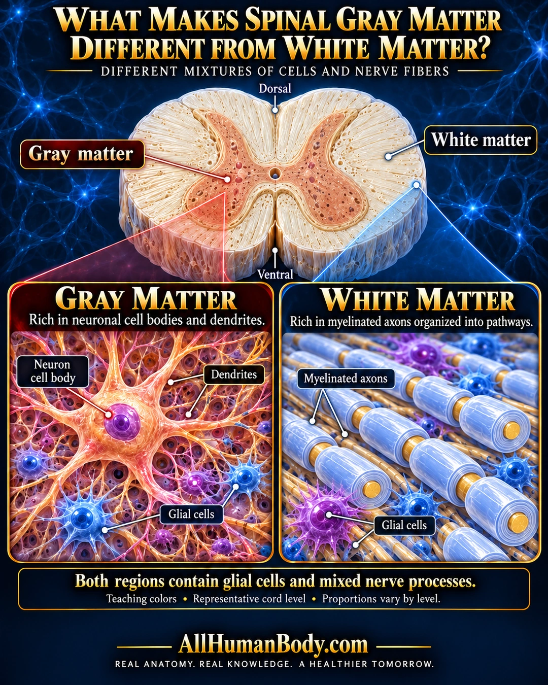
Spinal gray and white matter differ in their mixtures of cells and nerve processes. Central gray matter is rich in neuronal cell bodies and dendrites; surrounding white matter contains many myelinated axons, and both regions contain glial cells.
READ THE IMAGE
What the illustration shows
Navy and gold poster compares spinal gray and white matter. A short transverse cord segment has a central peach butterfly labeled gray matter and an ivory periphery labeled white matter, with dorsal above and ventral below. A red connector leads to a gray-matter panel with a large neuron cell body, branching dendrites and small glial cells. A blue connector from peripheral white matter leads to a panel of individual gold axons, each encircled by its own pale-blue myelin sheath, with glial cells between fibers. Text states that both regions contain glial cells and mixed nerve processes. Teaching colors and level variation are noted above the AllHumanBody.com brand and full slogan.
How to interpret this poster
- Teaching colors, cell shapes and representative proportions are not histology or MRI. Proportions vary with cord level; no lesion or pathway diagnosis.
- AI-assisted educational illustration; not physician certification, microscopy, a diagnosis or individualized medical advice.
- Fine annotations need enlargement; the full description provides an accessible text alternative.
- Display the full uncropped illustration; do not trim titles, legends or boundary annotations.
AI-assisted educational illustration, adapted into web-sized images without changing its composition. This is general education, not an individual diagnosis or treatment recommendation.
EVIDENCE / CONTEXT
Sources and what they support
- UTHealth Neuroscience Online, Anatomy of the Spinal Cord, sections3.4 and3.7 ↗
Central butterfly gray matter, cellular populations and longitudinal white-matter fibers.
- University of Leeds Histology Guide ↗
Central/peripheral arrangement and comparative cell/axon-rich microanatomy; do not repeat literal simplification that white matter has no cell bodies, because glial cells are present.