Nervous / VISUAL EXPLAINER
What Is the Partition Between Your Brain’s Hemispheres?
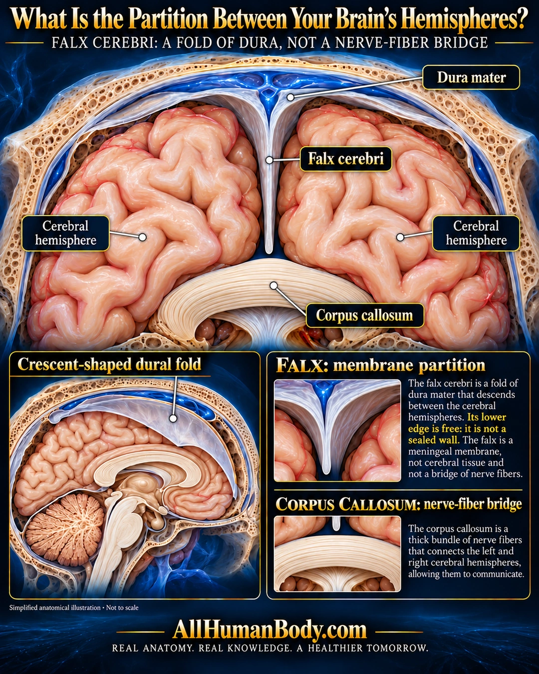
The falx cerebri is a crescent-shaped fold of dura that descends between the cerebral hemispheres. It is a membrane partition, distinct from the corpus callosum’s nerve-fiber connection between the two sides.
READ THE IMAGE
What the illustration shows
English anatomical poster on a navy field. A front-facing opened cranium shows pale peach cerebral hemispheres on either side of a thin ivory falx continuous with cranial dura. The falx ends freely above a separate ivory corpus callosum bridge. Labels identify dura, falx, cerebral hemisphere and corpus callosum. A sagittal inset shows the crescent-shaped dural sheet. Two close-ups contrast the free membrane partition with the nerve-fiber bridge. Gold AllHumanBody.com and the full project slogan appear at the bottom.
How to interpret this poster
- Simplified dural relationships, not a sealed wall or proof of cortical adhesion. No hemorrhage boundary, tumor, imaging or surgical inference.
- AI-assisted educational illustration; not physician certification, microscopy, a diagnosis or individualized medical advice.
- Fine annotations need enlargement; the full description provides an accessible text alternative.
- Display the full uncropped illustration; do not trim titles, legends or boundary annotations.
AI-assisted educational illustration, adapted into web-sized images without changing its composition. This is general education, not an individual diagnosis or treatment recommendation.
EVIDENCE / CONTEXT
Sources and what they support
- Manipal Academy of Higher Education anatomy specimen teaching description ↗
Dural fold identity, downward midline position, anterior and posterior attachments; venous sinus context.
- Cleveland Clinic medically reviewed anatomy guide ↗
Interhemispheric fiber connection, axonal white matter, and curved profile.