Nervous / VISUAL EXPLAINER
Where Is the Blood–Brain Barrier Built?
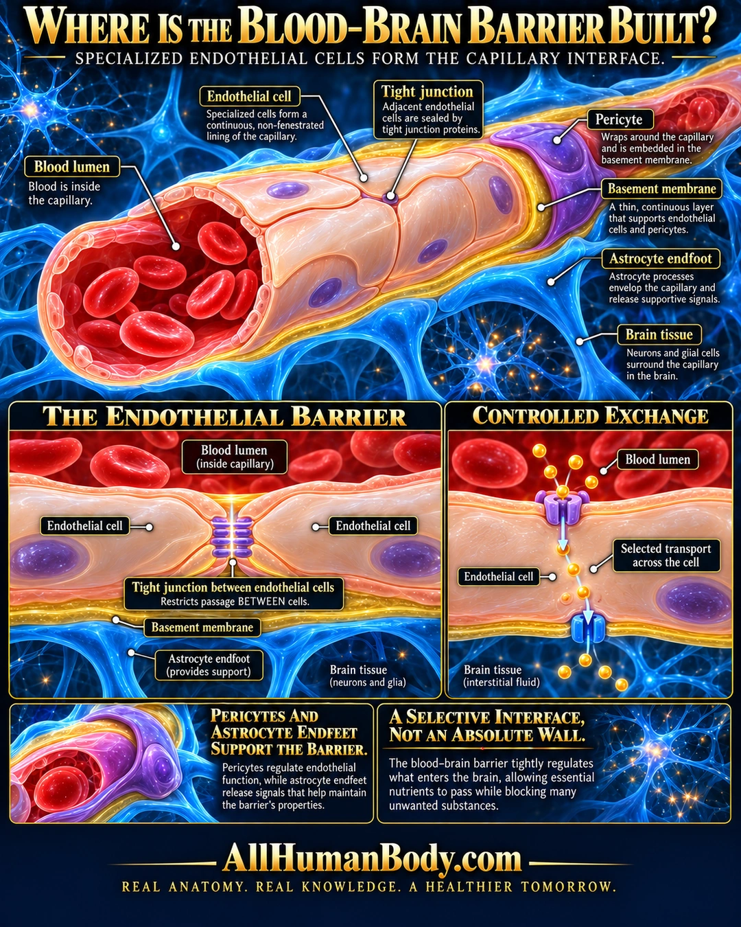
Specialized endothelial cells form the interface of typical blood–brain barrier capillaries. Tight junctions restrict passage between cells, while selective transport supplies neural tissue. Pericytes and astrocyte endfeet support this endothelial barrier.
READ THE IMAGE
What the illustration shows
Navy-and-gold poster shows a brain capillary cutaway with red blood cells inside, pale peach endothelial cells, gold basement membrane, purple pericyte and blue astrocyte endfeet outside. Lower-left inset shows a tight junction between two endothelial cells. Lower-right inset shows selected transport through one cell, crossing its upper and lower membranes with cytoplasm between. Text calls the barrier selective, not absolute. Gold brand and full slogan sit on plain navy footer.
How to interpret this poster
- Typical barrier capillary, not every CNS vessel or circumventricular region. Selective does not mean absolutely impermeable.
- Endothelial tiles, nuclei, basement-membrane thickness, pericyte embedding and endfoot coverage are illustrative, not electron microscopy.
- The two-membrane route is not a named transporter, substrate chemistry or protein count. No universal zero-vesicle claim or drug-delivery/barrier-opening advice.
- AI-assisted educational illustration; not physician certification, microscopy, a diagnosis or individualized medical advice.
- Fine annotations need enlargement; the full description provides an accessible text alternative.
- Display the full uncropped illustration; do not trim titles, legends or boundary annotations.
AI-assisted educational illustration, adapted into web-sized images without changing its composition. This is general education, not an individual diagnosis or treatment recommendation.
EVIDENCE / CONTEXT
Sources and what they support
- Neuroscience Online: Blood Brain Barrier and Cerebral Metabolism ↗
Endothelial interface, junctional restriction, adjacent support cells and selective transport.