Nervous / VISUAL EXPLAINER
Why Is a Sensory Neuron’s Cell Body Beside Its Main Route?
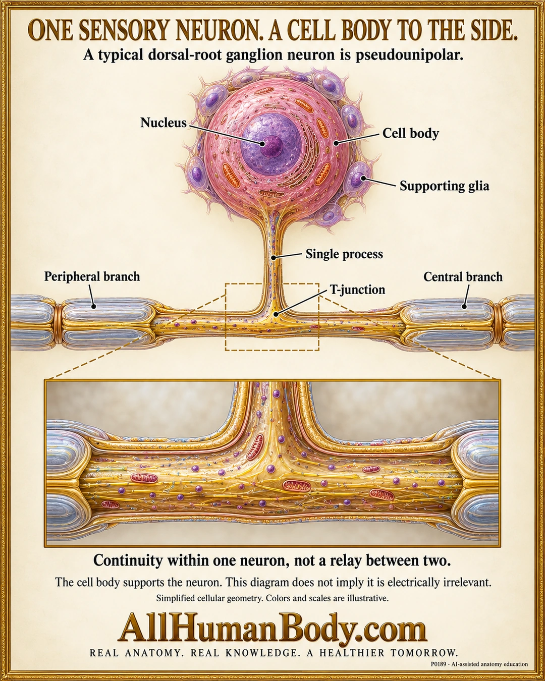
A typical dorsal-root ganglion neuron is pseudounipolar: one process from its cell body divides into peripheral and central branches. This is continuity within one neuron, not a relay between two neurons, and the cell body remains part of its function.
READ THE IMAGE
What the illustration shows
Ivory-and-gold anatomy poster showing one pink round sensory-neuron cell body with a purple nucleus and surrounding supporting glia. A single gold process descends to a continuous T-shaped axon, with peripheral branch left and central branch right, each with pale myelin sleeves. A larger lower cutaway shows continuous cytoplasm through the junction without a synaptic cleft. Text explains pseudounipolar geometry and that the cell body is not electrically irrelevant. Colors, scales, organelles and sheath thickness are illustrative. Gold AllHumanBody.com and the full brand slogan appear below.
How to interpret this poster
- Not every sensory neuron has this geometry. The drawing does not imply electrical irrelevance of the soma or that signals can never invade it.
- Organelles, glial shapes, sheath lamellae, spacing and scales are conceptual, not measured ultrastructure.
- Supporting satellite glia are distinct from skeletal-muscle satellite cells. No pain, disease or fixed cell-count claim.
- AI-assisted educational illustration; not physician certification, microscopy, a diagnosis or individualized medical advice.
- Fine annotations need enlargement; the full description provides an accessible text alternative.
- Display the full uncropped illustration; do not trim titles, legends or boundary annotations.
AI-assisted educational illustration, adapted into web-sized images without changing its composition. This is general education, not an individual diagnosis or treatment recommendation.
EVIDENCE / CONTEXT
Sources and what they support
- NLM MeSH: Ganglia, Spinal ↗
Dorsal-root ganglion sensory-neuron single process bifurcation into peripheral/central branches.
- University of Leeds Histology: Nerves and ganglia ↗
Sensory neuronal cell bodies and supporting glia in ganglia.