Nervous / VISUAL EXPLAINER
Where Does an Autonomic Signal Change Neurons?
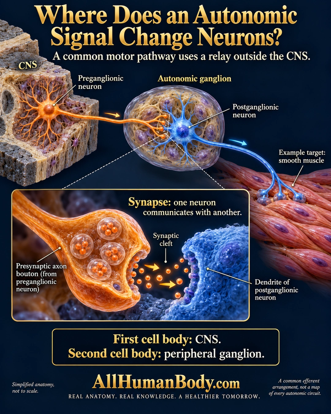
A common autonomic motor route uses two neurons: one has its cell body in the central nervous system and communicates in a peripheral ganglion with another neuron that sends an axon toward a visceral target.
READ THE IMAGE
What the illustration shows
English poster titled Where Does an Autonomic Signal Change Neurons? An orange preganglionic neuron has its cell body inside a CNS tissue block. Its axon ends on a separate blue neuron inside an autonomic ganglion. The blue postganglionic axon continues toward an example smooth-muscle target. A large inset shows an orange axon bouton, a synaptic cleft and a blue dendrite, with directional arrows across the gap. Text contrasts the first CNS cell body with the second peripheral cell body, and calls this a common arrangement rather than every autonomic circuit. Gold AllHumanBody.com and the full project slogan are intact.
How to interpret this poster
- Common efferent arrangement, not every autonomic or involuntary circuit; adrenal and enteric organization have important differences.
- Ganglion cell density, synaptic shapes and terminal field are schematic rather than verified microscopy.
- No transmitter inventory, universal opposing dual supply, fixed organ effect or therapeutic vagus claim.
- AI-assisted educational illustration; not physician certification, microscopy, a diagnosis or individualized medical advice.
- Fine annotations need enlargement; the full description provides an accessible text alternative.
- Display the full uncropped illustration; do not trim titles, legends or boundary annotations.
AI-assisted educational illustration, adapted into web-sized images without changing its composition. This is general education, not an individual diagnosis or treatment recommendation.
EVIDENCE / CONTEXT
Sources and what they support
- Anatomy and Physiology 2e: 15.1 Divisions of the Autonomic Nervous System ↗
Central/peripheral soma placement, ganglionic relay, targets and exceptions; preserved direct selected lines 41–60 and 68–75.
- University of Minnesota: Autonomic Nervous System — General Features ↗
Two-neuron visceral efferent organization.