Nervous / VISUAL EXPLAINER
Where Does Cerebrospinal Fluid Come From?
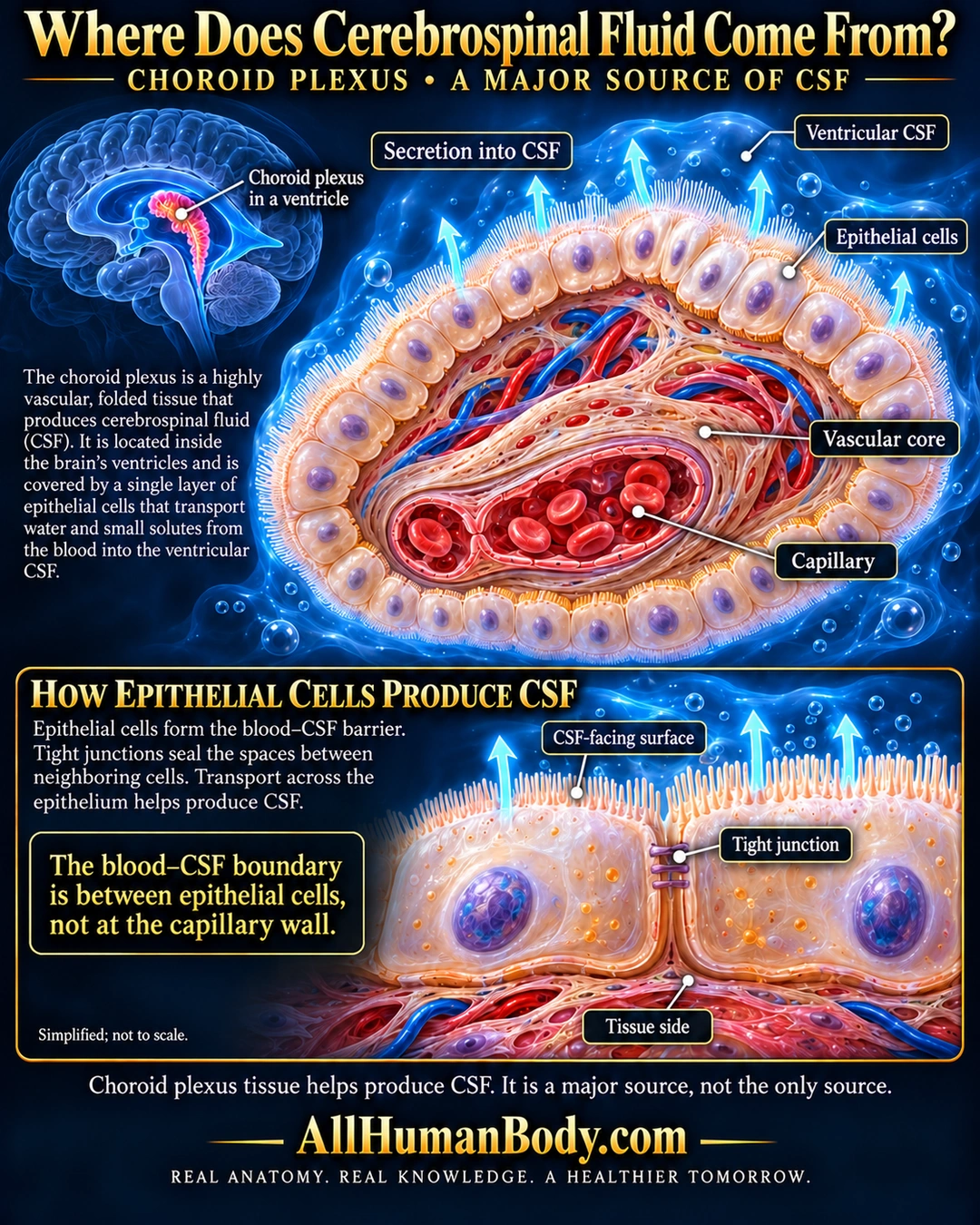
The choroid plexus projects into brain ventricles and is a major source of cerebrospinal fluid. An epithelial layer surrounds a vascular core; epithelial tight junctions help form the blood–CSF barrier while regulated transport across the layer contributes to secretion.
READ THE IMAGE
What the illustration shows
Navy-and-gold medical poster titled Where Does Cerebrospinal Fluid Come From? A small blue brain locator points to the choroid plexus. A large cutaway shows a pale epithelial coat surrounding a vascular core containing red blood cells within a capillary. Cyan arrows point outward into ventricular CSF. A lower close-up labels the CSF-facing surface, tight junction between neighboring epithelial cells, and tissue side. Text identifies the plexus as a major, not the only, CSF source. Gold AllHumanBody.com branding and the full slogan appear below.
How to interpret this poster
- Major source does not mean all CSF, and no fixed production percentage is assigned. Water is not described as actively pumped.
- The barrier callout concerns epithelial paracellular sealing; cell membranes also mediate selective transcellular transport.
- Surface projections, junction bars, vessel diameter and tissue thickness are schematic. No pressure diagnosis, caffeine advice, glymphatic detox or therapeutic promise.
- AI-assisted educational illustration; not physician certification, microscopy, a diagnosis or individualized medical advice.
- Fine annotations need enlargement; the full description provides an accessible text alternative.
- Display the full uncropped illustration; do not trim titles, legends or boundary annotations.
AI-assisted educational illustration, adapted into web-sized images without changing its composition. This is general education, not an individual diagnosis or treatment recommendation.
EVIDENCE / CONTEXT
Sources and what they support
- University of Basel Histology: Choroid plexus ↗
Human specimen description of ventricular epithelial layer, vascular core and epithelial tight junctions.
- Development and functions of the choroid plexus-cerebrospinal fluid system ↗
Principal CSF-source role and adult tissue organization; abstract and Figure1 caption, not full article.
Evidence correspondence is limited to the abstract.