Urinary / VISUAL EXPLAINER
How Does the Bladder Lining Change Shape as It Fills?
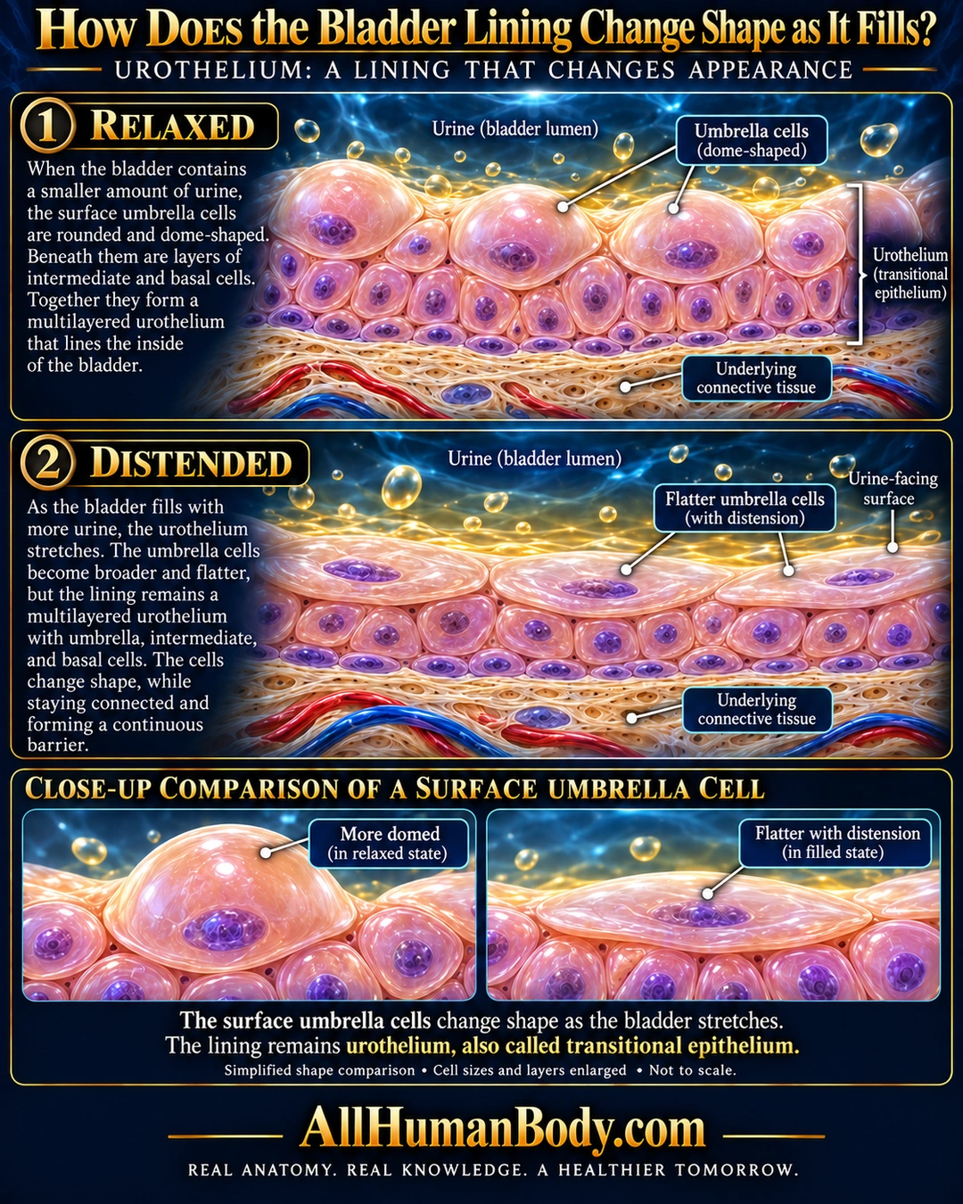
The bladder’s urothelial lining changes appearance as it stretches. Its surface umbrella cells become flatter with distension while remaining part of a continuous urothelium.
READ THE IMAGE
What the illustration shows
English poster on navy with relaxed and distended urothelial strips. Rounded peach umbrella cells cap the top strip; broader flatter surface cells cap the second. Purple nuclei and underlying smaller cells remain visible, with continuous epithelial tissue above pale fibrous connective tissue and red/blue vessels. A bracket identifies the entire urothelium. Lower closeups compare domed and flatter cells. Gold droplets above the surfaces are illustrative fluid symbols, not measured molecules or gas. Footer states the lining remains urothelium and that sizes/layers are enlarged, not to scale. Gold AllHumanBody.com and complete slogan appear below.
How to interpret this poster
- No fixed layer count, filling volume, strain or timing. Shape change is not conversion to squamous epithelium or evidence of cell loss.
- AI-assisted educational illustration; not physician certification, microscopy, a diagnosis or individualized medical advice.
- Fine annotations need enlargement; the full description provides an accessible text alternative.
- Display the full uncropped illustration; do not trim titles, legends or boundary annotations.
AI-assisted educational illustration, adapted into web-sized images without changing its composition. This is general education, not an individual diagnosis or treatment recommendation.
EVIDENCE / CONTEXT
Sources and what they support
- University of Minnesota Histology Guide ↗
Urothelium and dome-shaped versus flattened umbrella cells.
- University of Leeds histology ↗
Distended urothelium looks flatter; folded mucosa accommodates volume changes.