Musculoskeletal / VISUAL EXPLAINER
Where Do Muscle Satellite Cells Live?
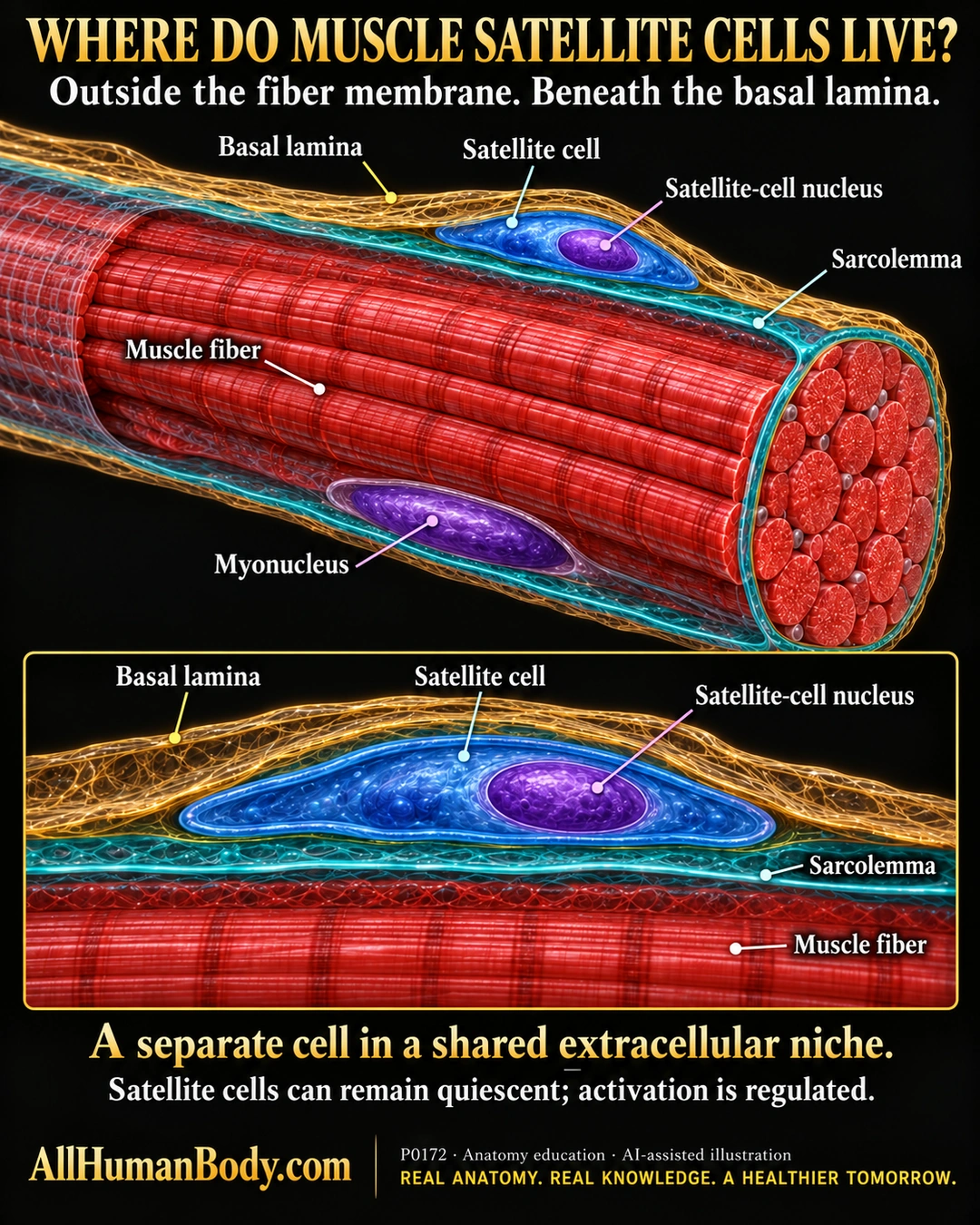
A skeletal-muscle satellite cell is a separate cell beside the muscle fiber, between its plasma membrane and basal lamina. Satellite cells can remain quiescent; activation and self-renewal depend on regulated interactions rather than continuous automatic division.
READ THE IMAGE
What the illustration shows
Black-and-gold poster titled Where Do Muscle Satellite Cells Live? A dimensional red skeletal muscle fiber contains a purple myonucleus beneath a teal sarcolemma. A separate blue satellite cell with its own purple nucleus lies above that membrane and below a gold mesh representing basal lamina. A lower magnified panel repeats this outside-membrane, beneath-lamina relationship, with labels for the satellite cell, its nucleus, sarcolemma, basal lamina and muscle fiber. Text states that activation is regulated. Gold AllHumanBody.com branding and the complete slogan appear along the bottom. Colors and thicknesses are illustrative, not measured.
How to interpret this poster
- Basal lamina is extracellular matrix, not another cell membrane; mesh texture, membrane thickness and relative sizes are exaggerated.
- The example myonucleus does not imply one nucleus per skeletal-muscle fiber.
- Research review includes experimental systems, not proof of human treatment benefits, muscle-memory permanence, hypertrophy guarantees or repair timelines.
- AI-assisted educational illustration; not physician certification, microscopy, a diagnosis or individualized medical advice.
- Fine annotations need enlargement; the full description provides an accessible text alternative.
- Display the full uncropped illustration; do not trim titles, legends or boundary annotations.
AI-assisted educational illustration, adapted into web-sized images without changing its composition. This is general education, not an individual diagnosis or treatment recommendation.
EVIDENCE / CONTEXT
Sources and what they support
- Satellite cells and the muscle stem cell niche ↗
Separate sublaminar cell location, quiescence and regulated activation/self-renewal.