Musculoskeletal / VISUAL EXPLAINER
How Does an Electrical Signal Reach Deep Inside a Muscle Fiber?
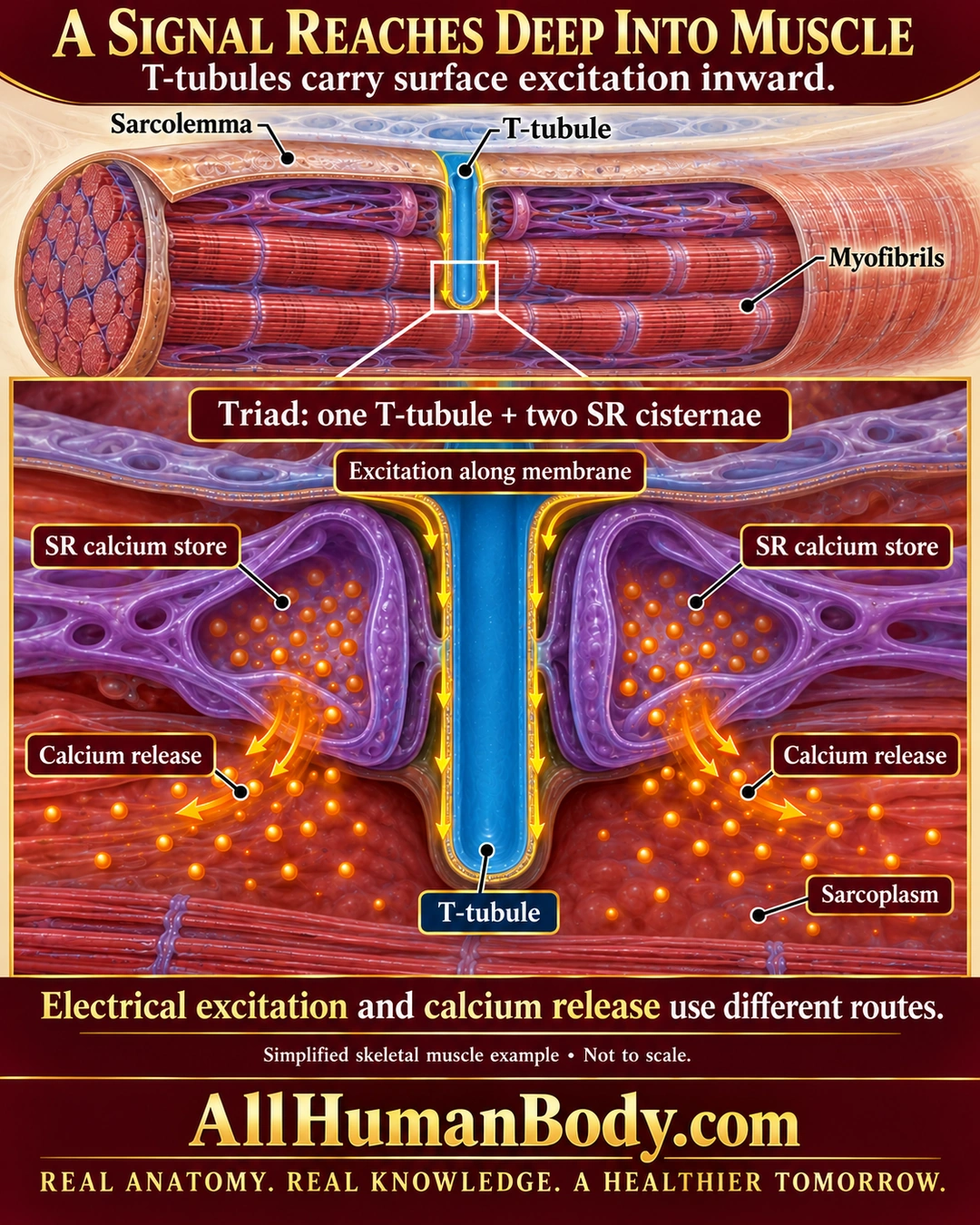
T-tubules extend the skeletal muscle cell surface inward. At a triad, one tubule lies beside two sarcoplasmic-reticulum cisternae; excitation along the tubule membrane couples to calcium release from the separate internal store into sarcoplasm.
READ THE IMAGE
What the illustration shows
Dimensional cutaway of a skeletal muscle fiber with labeled sarcolemma, blue inward T-tubule and red myofibrils. An enlargement shows the blue tubule between two separate purple SR calcium stores. Yellow arrows follow the tubule membrane; orange calcium dots and arrows leave SR into the surrounding sarcoplasm. Text distinguishes excitation from calcium release and identifies one tubule plus two cisternae as a triad. Gold AllHumanBody.com branding and the full slogan appear below. Simplified, not to scale.
How to interpret this poster
- Selected local invagination, not the complete transverse network or molecular channel reconstruction.
- Electrical excitation and calcium release use separate routes; the tubule and SR lumens are not fused.
- No measured timing, cardiac-style dominant external calcium entry or treatment claim.
- AI-assisted educational illustration; not physician certification, microscopy, a diagnosis or individualized medical advice.
- Fine annotations need enlargement; the full description provides an accessible text alternative.
- Display the full uncropped illustration; do not trim titles, legends or boundary annotations.
AI-assisted educational illustration, adapted into web-sized images without changing its composition. This is general education, not an individual diagnosis or treatment recommendation.
EVIDENCE / CONTEXT
Sources and what they support
- Anatomy and Physiology 2e: 10.2 Skeletal Muscle ↗
Surface invagination, triad and SR calcium release; preserved direct excitation-contraction section and production reread.