Musculoskeletal / VISUAL EXPLAINER
Are All Your Muscles Built the Same Way?
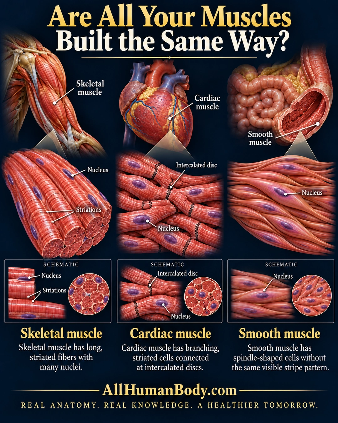
Muscle tissues have different cellular architecture. Skeletal fibers are long, striated and multinucleate; cardiac cells branch and connect at intercalated discs; smooth cells taper without the same visible transverse stripe pattern.
READ THE IMAGE
What the illustration shows
Three-column anatomy comparison of skeletal, cardiac and smooth muscle. An upper-arm muscle, heart and intestinal-wall cutaway locate the tissues. Below, skeletal fibers are long and striped with peripheral nuclei; cardiac cells form a branching striped network with central nuclei and dark intercalated junctions; smooth cells are tapered overlapping spindles without transverse stripes. Lower schematic closeups repeat nuclei and junction details. Each column has a concise explanation, with gold AllHumanBody.com branding at the bottom.
How to interpret this poster
- Illustrative magnifications are not identical measured cell sizes or diagnostic microscopy.
- Representative nuclei and organ locators do not map all tissue variation.
- No claim that skeletal activity is always conscious or that the heart cannot fatigue.
- AI-assisted educational illustration; not physician certification, microscopy, a diagnosis or individualized medical advice.
- Fine annotations need enlargement; the full description provides an accessible text alternative.
- Display the full uncropped illustration; do not trim titles, legends or boundary annotations.
AI-assisted educational illustration, adapted into web-sized images without changing its composition. This is general education, not an individual diagnosis or treatment recommendation.
EVIDENCE / CONTEXT
Sources and what they support
- Anatomy and Physiology 2e: 4.4 Muscle Tissue and Motion ↗
Comparison of skeletal, cardiac and smooth cellular morphology.
- SEER Training: Types of Muscle Tissue ↗
Normal tissue shapes and representative locations.