Cardiovascular / VISUAL EXPLAINER
How Do Heart Muscle Cells Stay Joined While Sharing Signals?
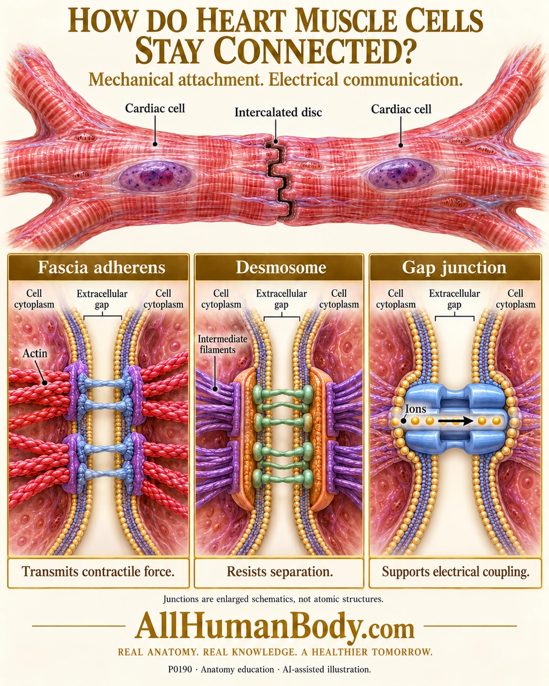
Intercalated discs join neighboring cardiac muscle cells through multiple junction systems. Adhesion junctions transmit force and help resist separation, while gap junctions allow electrical communication between cell interiors.
READ THE IMAGE
What the illustration shows
Ivory and gold poster shows two branching, striated cardiac cells with purple nuclei meeting at a stepped intercalated disc. Three enlarged panels below show fascia adherens anchoring red actin at paired membranes, a desmosome anchoring purple intermediate filaments, and a blue docked gap-junction channel carrying gold ions horizontally from one cell interior to the other. Captions distinguish force transmission, resistance to separation and electrical coupling. Gold AllHumanBody.com and full slogan appear at bottom.
How to interpret this poster
- Molecular shapes, membrane gaps, junction sizes and striations are schematic, not atomic structures, protein counts or sarcomere measurements.
- A passage arrow does not make a gap junction obligatorily one-way. Discs contain more than desmosomes alone.
- No ECG, rhythm diagnosis, heart-rate control or treatment claim.
- AI-assisted educational illustration; not physician certification, microscopy, a diagnosis or individualized medical advice.
- Fine annotations need enlargement; the full description provides an accessible text alternative.
- Display the full uncropped illustration; do not trim titles, legends or boundary annotations.
AI-assisted educational illustration, adapted into web-sized images without changing its composition. This is general education, not an individual diagnosis or treatment recommendation.
EVIDENCE / CONTEXT
Sources and what they support
- University of Leeds Histology: Muscle cell junctions ↗
Fascia adherens, desmosomal anchorage and gap-junction distinctions.
- Anatomy and Physiology 2e: 10.7 Cardiac Muscle Tissue ↗
Cardiac cell mechanical/electrical coupling; preserved directly read text, not sole source of ultrastructure.