Musculoskeletal / VISUAL EXPLAINER
What Actually Shortens Inside a Muscle?
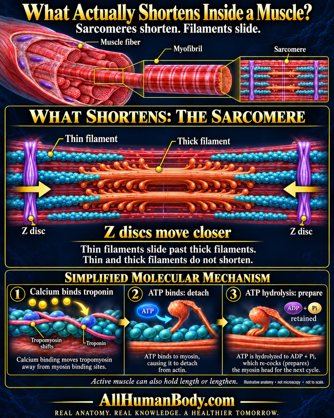
During muscle shortening, sarcomere boundaries approach as thin and thick filaments slide into greater overlap. The filaments do not themselves shorten; calcium regulates access and ATP supports myosin cycling.
READ THE IMAGE
What the illustration shows
Navy-and-gold educational poster titled “What Actually Shortens Inside a Muscle?” A cutaway muscle fiber is enlarged to show a myofibril and sarcomere. The main diagram shows one sarcomere: teal thin filaments extend inward from purple Z discs and overlap copper thick filaments. Yellow arrows at the two Z discs point toward the center. Text explains that Z discs move closer, thin filaments slide past thick filaments, and neither filament type shortens. Three simplified molecular panels show calcium binding troponin and shifting tropomyosin, ATP binding myosin to permit detachment, and ATP hydrolysis preparing the head with ADP and phosphate retained. A note says active muscle can also hold length or lengthen. Footer: AllHumanBody.com; REAL ANATOMY. REAL KNOWLEDGE. A HEALTHIER TOMORROW. The illustration is labeled not microscopy and not to scale.
How to interpret this poster
- The final poster has one qualitative sarcomere schematic, not a measured relaxed-versus-shortened comparison.
- Active muscle can also hold length or lengthen.
- Molecular shapes and retained reaction products are schematic, not microscopy or a complete cross-bridge cycle.
- AI-assisted educational illustration; not physician certification, microscopy, a diagnosis or individualized medical advice.
- Fine annotations need enlargement; the full description provides an accessible text alternative.
AI-assisted educational illustration, adapted into web-sized images without changing its composition. This is general education, not an individual diagnosis or treatment recommendation.
EVIDENCE / CONTEXT
Sources and what they support
- Anatomy and Physiology 2e: 10.3 Muscle Fiber Contraction and Relaxation ↗
Sliding filaments, calcium–troponin regulation, ATP-dependent detachment and myosin preparation.