Integumentary / VISUAL EXPLAINER
Why Can Veins Look Blue Through Skin?
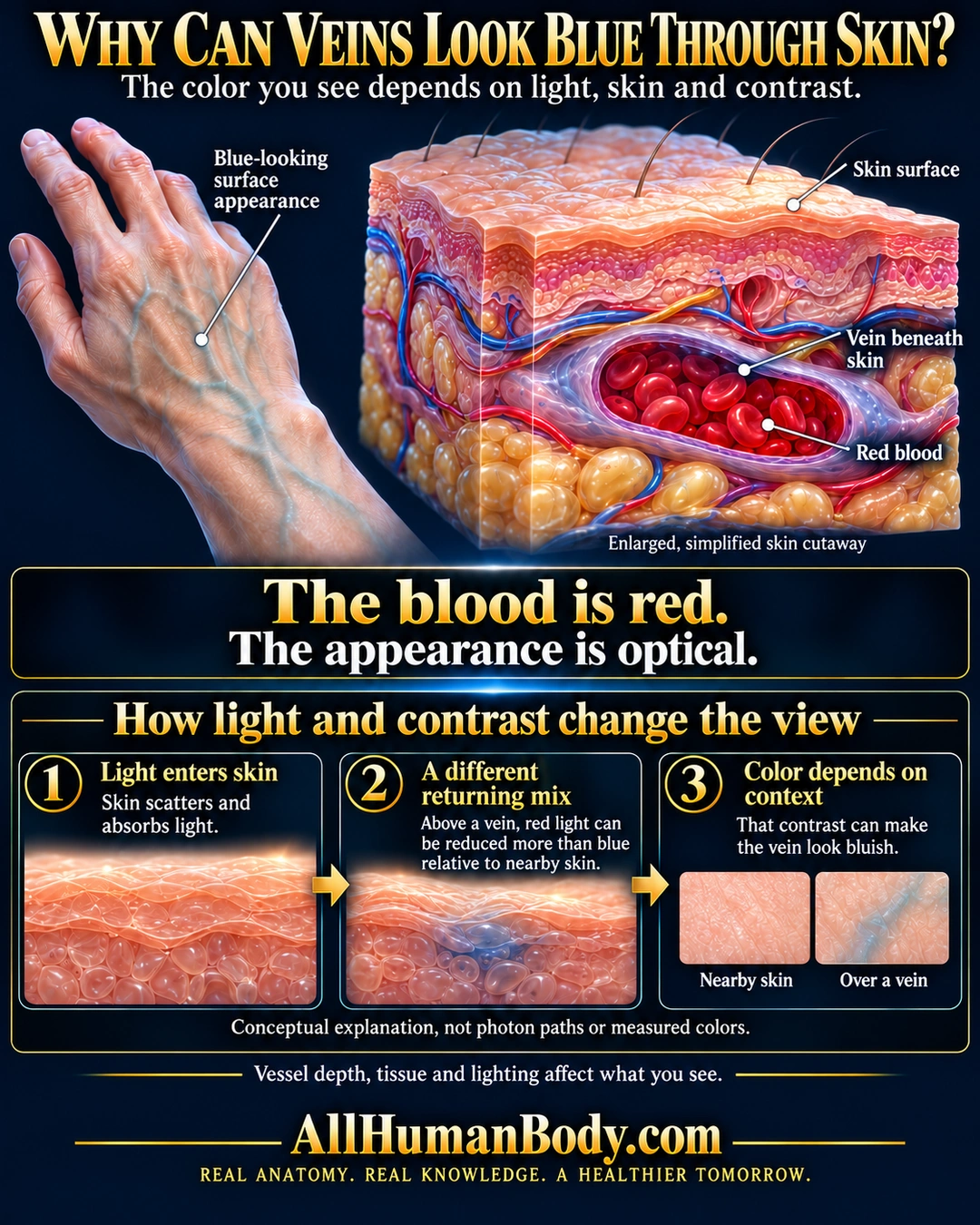
Veins may look blue beneath skin even though the blood inside is red. Light interacting with skin and blood changes the color seen relative to nearby skin.
READ THE IMAGE
What the illustration shows
English poster titled “Why Can Veins Look Blue Through Skin?” A five-fingered hand and forearm at upper left show subtle blue-green vessel-like lines through intact peach skin, labeled blue-looking surface appearance. To the right, an enlarged skin block has surface hairs, layered tissue, small red and blue vessels, yellow cord-like structures and yellow fat. A cutaway of a larger purple-gray vessel below the skin exposes dark red blood and bright red cells. Leaders identify the skin surface, vein beneath skin and red blood. Large central text says the blood is red and the appearance is optical. Three lower conceptual cards are joined by two rightward gold arrows: light enters skin; a different returning mix; color depends on context. The second card has blue shading beneath a modeled skin surface and states that red light can be reduced more than blue relative to nearby skin. The third compares nearby-skin and over-vein swatches. A note explicitly says this is a conceptual explanation, not photon paths or measured colors. A final caveat names vessel depth, tissue and lighting. Gold AllHumanBody.com and the full project slogan sit on a flat navy footer.
How to interpret this poster
- Color depends on depth, skin, lighting and perception. Panels are not measured photon trajectories; phantom findings are not universal across skin tones.
- AI-assisted educational illustration; not physician certification, microscopy, a diagnosis or individualized medical advice.
- Fine annotations need enlargement; the full description provides an accessible text alternative.
- Display the full uncropped illustration; do not trim titles, legends or boundary annotations.
AI-assisted educational illustration, adapted into web-sized images without changing its composition. This is general education, not an individual diagnosis or treatment recommendation.
EVIDENCE / CONTEXT
Sources and what they support
- Kienle et al., primary optical measurement/modeling study, Applied Optics, 1996; author-institution PDF ↗
Human-vein observation, scattering-medium experiments and simulation support wavelength-dependent tissue optics plus surrounding-color comparison. No figure, numerical depth threshold or layout is reused.
Evidence correspondence is limited to the abstract. - Wagner et al., primary optical phantom study, Biomedical Optics Express, 2026 ↗
Measured skin-like phantoms corroborate relative red-reflectance loss and contrast. Model lacks full layered-skin optics and uses a limited skin-color context, so it is not universal validation across skin tones.
- OpenStax Anatomy and Physiology 2e, authored textbook ↗
Vessels lie below the avascular epidermis; guides original cutaway compartment placement.