Integumentary / VISUAL EXPLAINER
Why Does Vitiligo Leave Pale Skin Patches?
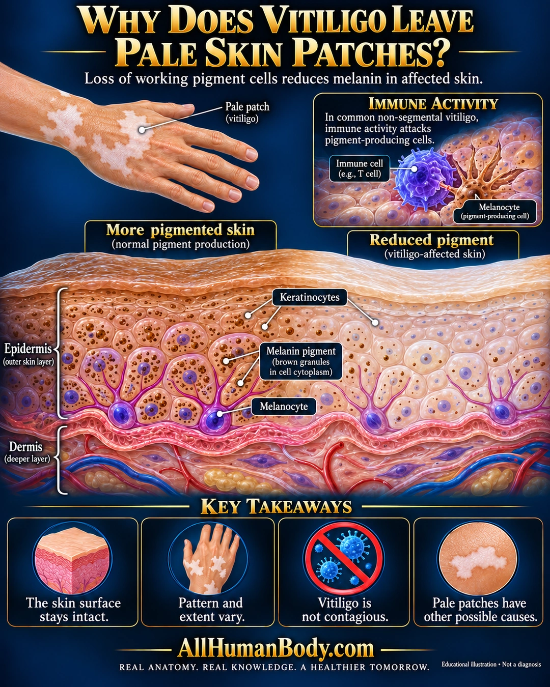
Loss of working pigment-producing cells reduces melanin in affected skin. In common non-segmental vitiligo, immune activity targets these cells. Pattern and extent vary, and vitiligo is not contagious.
READ THE IMAGE
What the illustration shows
Educational poster titled “Why Does Vitiligo Leave Pale Skin Patches?” A dorsal hand has pale patches. The main skin cross-section shows an intact epidermis across a more-pigmented left side and reduced-pigment right side. Branched melanocytes sit along the basal boundary; brown pigment granules lie in keratinocyte cytoplasm around purple nuclei, with fewer granules on the right. An inset illustrates an immune cell next to a melanocyte and limits the explanation to common non-segmental vitiligo. Four takeaways say the surface stays intact, pattern and extent vary, vitiligo is not contagious, and pale patches have other possible causes. Gold AllHumanBody.com branding on a navy field.
How to interpret this poster
- Not every pale patch or vitiligo subtype; intact-surface schematic is not diagnosis. Immune contact and cell numbers conceptual, no treatment or racial categories.
- AI-assisted educational illustration; not physician certification, a diagnosis or individualized medical advice.
- Fine annotations require enlargement; preserve the full uncropped illustration and its qualifiers.
AI-assisted educational illustration, adapted into web-sized images without changing its composition. This is general education, not an individual diagnosis or treatment recommendation.
EVIDENCE / CONTEXT
Sources and what they support
- NIAMS: Vitiligo ↗
Immune-associated pigment-cell loss.
- NHS: Vitiligo ↗
Functioning melanocytes, non-segmental scope and noncontagiousness.
- Perilesional T cells and vitiligo melanocytes ↗
T-cell targeting in patient-derived skin explants supports example label.
Evidence correspondence is limited to the abstract.