Integumentary / VISUAL EXPLAINER
How Your Skin Renews Its Surface
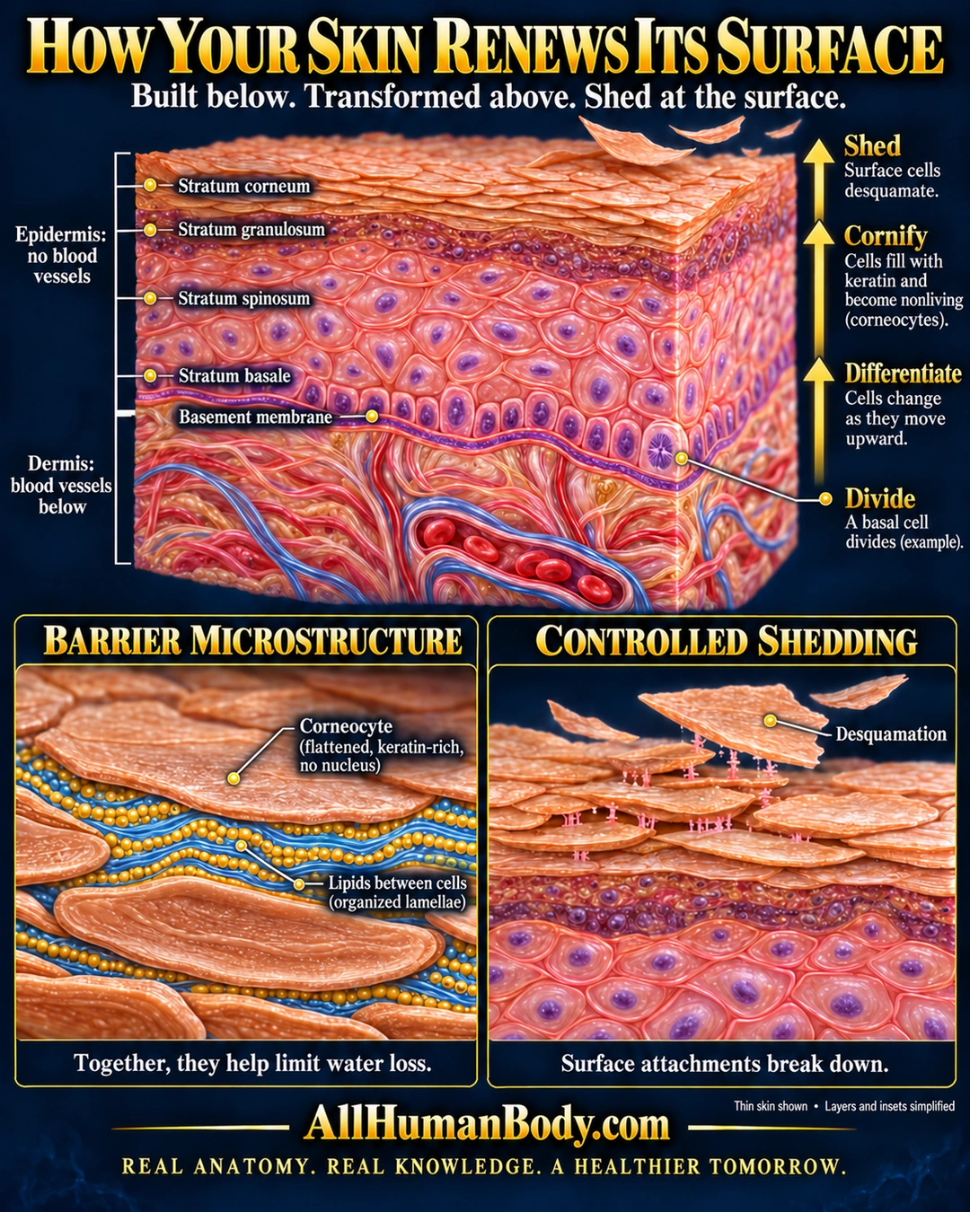
New epidermal cells are produced below the surface and change as they move outward. Flattened corneocytes and surrounding lipids contribute to the barrier while surface cells are gradually shed.
READ THE IMAGE
What the illustration shows
Illustrated poster titled “How Your Skin Renews Its Surface.” A cutaway of thin skin shows four epidermal layers, from the deepest stratum basale through the stratum spinosum and granulosum to flattened surface cells in the stratum corneum. A wavy basement membrane separates the epidermis from the connective-tissue dermis. Blood vessels appear only below this boundary. One basal cell is illustrated dividing. Upward arrows describe division, differentiation, cornification and shedding. A barrier inset shows flattened, keratin-rich corneocytes without nuclei, with organized lipid layers between them; together these help limit water loss. A shedding inset shows attachments between outer cells breaking down as superficial cells detach. Cell shapes, spacing, layer thicknesses and inset structures are simplified, and the region brackets are approximate. Fine labels are best read by zooming. Branded AllHumanBody.com.
How to interpret this poster
- Thin skin is shown; cell counts, layer thicknesses and region brackets are schematic.
- Renewal stages overlap, and the poster does not set a universal turnover clock.
- Shedding involves coordinated changes, not a single universal attachment-breaking event.
- AI-assisted educational illustration; not physician certification, microscopy, a diagnosis or individualized medical advice.
- Fine annotations need enlargement; the full description provides an accessible text alternative.
AI-assisted educational illustration, adapted into web-sized images without changing its composition. This is general education, not an individual diagnosis or treatment recommendation.
EVIDENCE / CONTEXT
Sources and what they support
- Anatomy and Physiology 2e: 5.1 Layers of the Skin ↗
Epidermal layer order, basal production, cornification and the distinction from vascular dermis.
- Cellular changes that accompany shedding of human corneocytes ↗
Human skin observations of changing cell attachments and extracellular lipid organization during shedding.
Evidence correspondence is limited to the abstract.