Integumentary / VISUAL EXPLAINER
Which Immune Cells Watch Between Your Skin Cells?
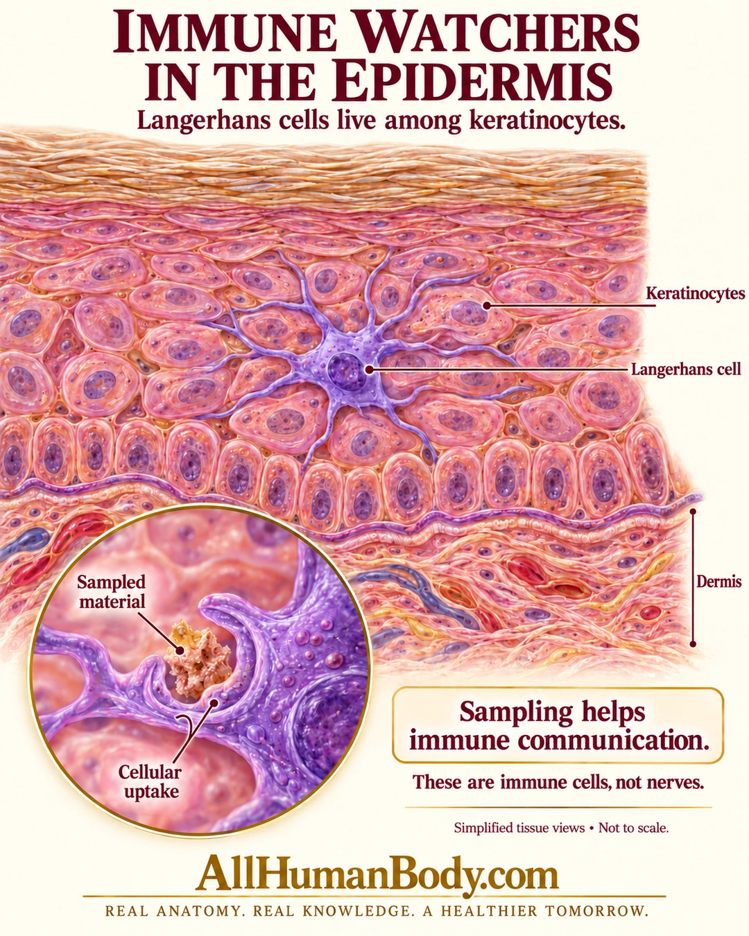
Langerhans cells are immune cells interspersed among epidermal keratinocytes. Their sampling and antigen-handling roles contribute to immune communication; a branching outline here does not represent a nerve.
READ THE IMAGE
What the illustration shows
An anatomy poster titled Immune Watchers in the Epidermis shows a purple branching Langerhans cell between pink keratinocytes, above the basal cell row and underlying dermis. Flattened surface layers appear above. Labels identify keratinocytes, the Langerhans cell and dermis. A circular close-up shows a purple cell taking up sampled material. Text says sampling helps immune communication and these are immune cells, not nerves. Simplified tissue views are not to scale. Gold AllHumanBody.com branding and the full slogan appear at the bottom.
How to interpret this poster
- Layer counts, colors, cell sizes and branches are schematic; identity cannot be diagnosed from color.
- No complete migration or antigen-presentation pathway is shown, and these are not the skin’s only defenses.
- No contested lineage shortcut, guaranteed immunity, rash diagnosis or treatment.
- AI-assisted educational illustration; not physician certification, microscopy, a diagnosis or individualized medical advice.
- Fine annotations need enlargement; the full description provides an accessible text alternative.
- Display the full uncropped illustration; do not trim titles, legends or boundary annotations.
AI-assisted educational illustration, adapted into web-sized images without changing its composition. This is general education, not an individual diagnosis or treatment recommendation.
EVIDENCE / CONTEXT
Sources and what they support
- Anatomy and Physiology 2e: 5.1 Layers of the Skin ↗
Spinous-layer location among keratinocytes and material uptake; preserved directly read paragraph and production reread, simplified macrophage taxonomy excluded.
- NLM MeSH: Langerhans Cells ↗
Epidermal antigen-presenting immune-cell identity.