Integumentary / VISUAL EXPLAINER
What Anchors Skin Cells to Their Foundation?
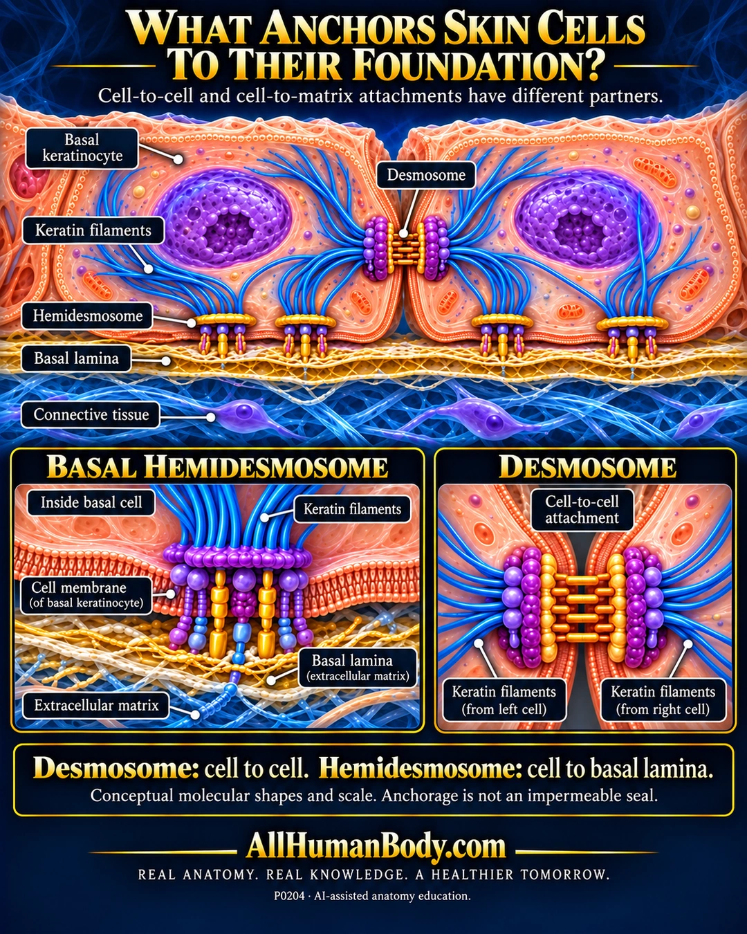
Desmosomes connect neighboring epithelial cells, while hemidesmosomes connect a basal cell to the underlying extracellular matrix. Their intracellular attachments link to keratin filaments, helping distribute mechanical stress.
READ THE IMAGE
What the illustration shows
English anatomy poster on a navy background with gold headings. Two peach basal keratinocytes have purple nuclei and blue keratin filaments. A desmosome bridges their facing lateral membranes. Hemidesmosomes sit along their basal surfaces above a gold basal lamina and blue connective tissue. Lower close-ups contrast a single basal cell attached to extracellular matrix with two neighboring cells joined by a desmosome. Labels identify keratin filaments, basal cell membrane and basal lamina. Text states that molecular shapes and scale are conceptual and anchorage is not an impermeable seal. The footer shows AllHumanBody.com and the slogan REAL ANATOMY. REAL KNOWLEDGE. A HEALTHIER TOMORROW.
How to interpret this poster
- Molecular shapes and spacing are schematic, not atomic structures. A hemidesmosome is not simply half of every desmosomal molecular component.
- This junction comparison is not a complete epidermal layer map, an impermeable seal or evidence of literal spikes on living cells.
- No blister diagnosis, wound treatment or skin-strength prediction.
- AI-assisted educational illustration; not physician certification, microscopy, a diagnosis or individualized medical advice.
- Fine annotations need enlargement; the full description provides an accessible text alternative.
- Display the full uncropped illustration; do not trim titles, legends or boundary annotations.
AI-assisted educational illustration, adapted into web-sized images without changing its composition. This is general education, not an individual diagnosis or treatment recommendation.
EVIDENCE / CONTEXT
Sources and what they support
- University of Leeds Histology: Desmosomes ↗
Cell-to-cell desmosomal and cell-to-matrix hemidesmosomal anchorage with intermediate filaments.
- University of Leeds Histology: Epidermal layers ↗
Keratinocyte junction and basal attachment context; same teaching institution, not independent experimental replication.