Integumentary / VISUAL EXPLAINER
What Does the Cuticle Connect at the Base of a Nail?
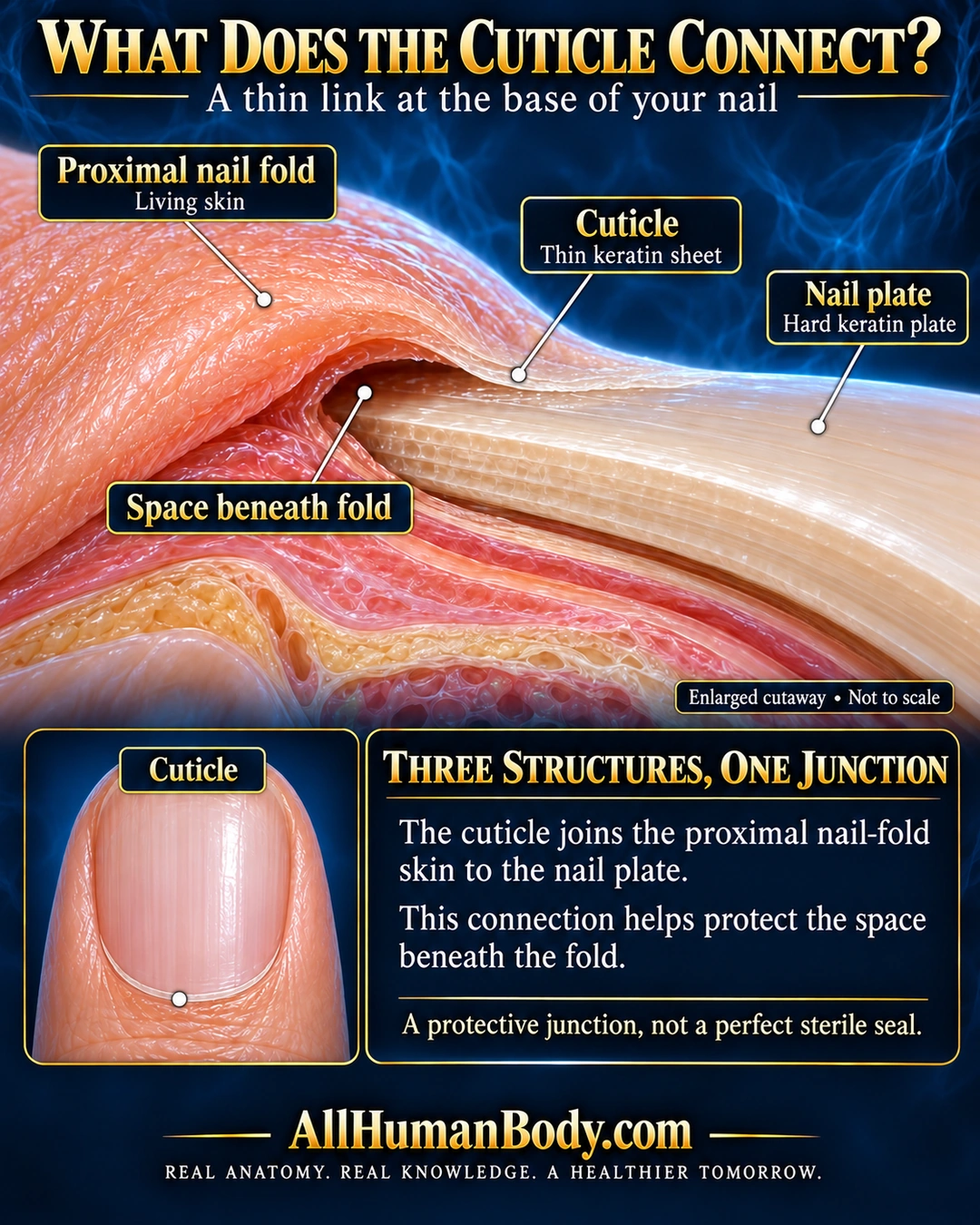
A thin keratin cuticle joins the skin of the proximal nail fold to the hard nail plate, helping protect the space beneath the fold. These are three distinct structures, not three names for one tissue.
READ THE IMAGE
What the illustration shows
English anatomy poster on a deep navy and cyan background titled What Does the Cuticle Connect? An enlarged cutaway shows a peach proximal nail fold at left arching over an ivory nail plate extending right. A very thin pale cuticle continues from the fold margin onto the plate. Four leaders identify living proximal nail fold, thin keratin cuticle, hard nail plate, and the small space beneath the fold above the plate. A lower-left top-view nail marks the cuticle at its bottom proximal border. A lower-right panel explains the connection and its protective role, with a note that it is not a perfect sterile seal. Gold AllHumanBody.com branding and the full slogan appear on the plain navy footer.
How to interpret this poster
- The recess and tissue thickness are enlarged, not a measured section or sterile seal. No manicure or infection prediction.
- AI-assisted educational illustration; not physician certification, microscopy, a diagnosis or individualized medical advice.
- Fine annotations need enlargement; the full description provides an accessible text alternative.
- Display the full uncropped illustration; do not trim titles, legends or boundary annotations.
AI-assisted educational illustration, adapted into web-sized images without changing its composition. This is general education, not an individual diagnosis or treatment recommendation.
EVIDENCE / CONTEXT
Sources and what they support
- DermNet dermatology teaching resource, lines503–505 ↗
Keratin sheet joins proximal nail-fold skin and nail plate and protects underlying space. Third claim is direct structural distinction, not a separate mechanistic discovery.