30-SECOND ORIENTATION / AN ILLUSTRATION AT ANOTHER SCALE
Connect the selected shape to normal anatomy.
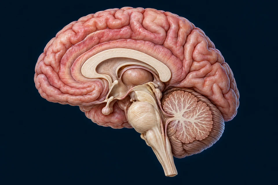
The image and editable labels explain anatomical relationships. They do not reveal new structures in the model you selected.
Generated gross anatomy, not MRI or microscopy. Anterior is left. Thin cyan rings indicate selected anatomical contexts, not nuclei boundaries, activation measurements or axon paths. Fine nuclei and fibre trajectories are not resolved. The hippocampus is not inserted into this midline view.
3-MINUTE UNDERSTANDING / ONE CANONICAL LEARNING PATH
Continue from what you can see to how it works.
Each route opens the corresponding existing lesson. The same deeper chapter remains the source of its full medical explanation.
Continue from gross anatomy to membrane and cellular mechanisms.
DEEPER LEARNING / ORIGINAL MODEL-LINKED EXPLANATIONS
Read the structure and function together.
External shape and internal organisation
The cerebrum occupies most of the cranial cavity. Behind the brainstem lies the cerebellum. Cortical gray matter and deeper nuclei connect through white-matter pathways; the external mesh alone does not reveal those internal relationships.
Networks rather than isolated boxes
Sensory processing, movement, learning and emotion depend on connected regions. A named lobe is a useful anatomical landmark, not a complete map of one exclusive function. The connected chapter distinguishes surface anatomy, deeper structures and mechanisms.
Protection and supply
The skull, meninges and cerebrospinal fluid support a controlled environment around the brain. Arterial supply delivers oxygen and substrates; the blood–brain barrier regulates exchange at the microvasculature. These relationships require cutaway and microscopic teaching views beyond this surface model.
Original AllHumanBody teaching explanations and retained chapter illustrations. The selected source-labelled model, authentic medical photographs and AI-assisted teaching illustrations are different resource types. This plate is an illustration, not a scan, microscopy or certification of hidden structures in the selected model. Existing chapter source and image-provenance records are retained.
The original source-linked notes above are preserved. Detailed mechanisms and their supporting references remain in the linked canonical chapter. Medical source checks do not constitute human medical review or personal diagnostic advice; fine generated anatomy and physical-phone acceptance are not independently certified.