30-SECOND ORIENTATION / AN ILLUSTRATION AT ANOTHER SCALE
Connect the selected shape to normal anatomy.
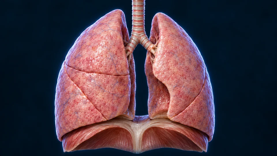
The image and editable labels explain anatomical relationships. They do not reveal new structures in the model you selected.
Retained AI-assisted gross-organ illustration, not a scan or an exact registration of the chosen mesh. Patient right is image left. This gross overview does not resolve microscopic airway layers, the exchange barrier or the neural control of breathing.Structure notes · further context
Trachea
Cartilage-supported passage divides into two main bronchi.
Right lung · three lobes
Image left: superior, middle and inferior lobes, separated by fissures.
Left lung · two lobes
Image right: superior and inferior lobes with a medial cardiac indentation.
Diaphragm context
Skeletal muscle beneath the lungs changes chest volume.
3-MINUTE UNDERSTANDING / ONE CANONICAL LEARNING PATH
Continue from what you can see to how it works.
Each route opens the corresponding existing lesson. The same deeper chapter remains the source of its full medical explanation.
Relate thoracic movement and recoil; lungs are not skeletal-muscle pumps.
DEEPER LEARNING / ORIGINAL MODEL-LINKED EXPLANATIONS
Read the structure and function together.
Position, lobes and airways
The right lung normally has three lobes. Branching airways enter at the hilum, and progressively smaller passages distribute inspired air toward alveoli.
Two flows meet across a barrier
Ventilation renews alveolar air. Pulmonary blood flow carries blood past that air, and gases diffuse along partial-pressure gradients across the alveolar–capillary barrier. Oxygen enters blood and carbon dioxide leaves it; the model surface cannot demonstrate this microscopic exchange.
Movement comes from the thorax
The diaphragm and other breathing muscles change thoracic volume. Pleural coupling allows the lung to follow chest movement. Lung tissue recoils during expiration; it does not pump by contracting like skeletal muscle.
Original AllHumanBody teaching explanations and retained chapter illustrations. The selected source-labelled model, authentic medical photographs and AI-assisted teaching illustrations are different resource types. This plate is an illustration, not a scan, microscopy or certification of hidden structures in the selected model. Existing chapter source and image-provenance records are retained.
The original source-linked notes above are preserved. Detailed mechanisms and their supporting references remain in the linked canonical chapter. Medical source checks do not constitute human medical review or personal diagnostic advice; fine generated anatomy and physical-phone acceptance are not independently certified.