1
Enter the kidney →
Locate cortex, medulla and the collecting route.
SELECTED ORGAN / STRUCTURE TO FUNCTION
A paired organ in the posterior abdomen links blood filtration to controlled water, electrolyte and acid–base balance.
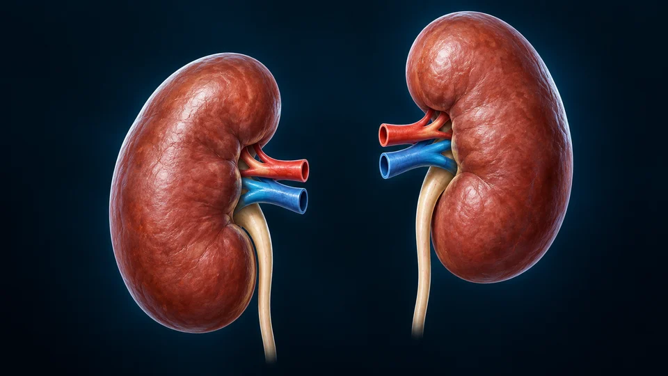
Both kidneys remain visible so that choosing one side does not silently mirror or rename the normal bilateral anatomy.
Read the connected anatomy →30-SECOND ORIENTATION / AN ILLUSTRATION AT ANOTHER SCALE
The image and editable labels explain anatomical relationships. They do not reveal new structures in the model you selected.
3-MINUTE UNDERSTANDING / ONE CANONICAL LEARNING PATH
Each route opens the corresponding existing lesson. The same deeper chapter remains the source of its full medical explanation.
1
Locate cortex, medulla and the collecting route.
2
Separate capillary blood from newly filtered fluid.
3
Connect tubular handling with physiological signals.
DEEPER LEARNING / ORIGINAL MODEL-LINKED EXPLANATIONS
The kidneys lie behind the peritoneal lining, on either side of the upper lumbar spine. The right is usually slightly lower. At the medial hilum, vessels enter and leave alongside the collecting pathway. Urine passes from the renal pelvis through the ureter to the bladder.
A glomerular capillary filter produces a fluid that enters the nephron tubule. Tubular cells then reclaim needed substances and secrete others. Collecting ducts adjust the final urine in response to physiological signals; urine is not simply blood with its cells removed.
The surface model establishes shape and sidedness. It does not expose a nephron, identify every vessel, or measure kidney function. Use the connected chapter for cortex, medulla, filtration and regulatory mechanisms.
Original AllHumanBody teaching explanations and retained chapter illustrations. The selected source-labelled model, authentic medical photographs and AI-assisted teaching illustrations are different resource types. This plate is an illustration, not a scan, microscopy or certification of hidden structures in the selected model. Existing chapter source and image-provenance records are retained.
The original source-linked notes above are preserved. Detailed mechanisms and their supporting references remain in the linked canonical chapter. Medical source checks do not constitute human medical review or personal diagnostic advice; fine generated anatomy and physical-phone acceptance are not independently certified.